Sialidosis
Sialidosis
Sialidosis is characterized by a deficiency of the digestive enzyme, alpha-neuraminidase. The lack of this enzyme results in an abnormal accumulation of complex carbohydrates known as mucopolysaccharides, and of fatty substances known as mucolipids. Both of these substances accumulate in bodily tissues. There are four types of Sialidosis: Each type of Sialidosis is characterized by the age of onset and by the type of physical and mental manifestations of this disorder. Type I affects both sexes with equal frequency.
For further details read the ‘History and Overview’ written for ISMRD in 2001. See also the 2018 review of Sialidosis prepared by Dr Sandra d’Azzo, a member of ISMRD’s professional advisory board. For frequently asked questions, read the ‘Sialidosis FAQ’.
How Common is it?
Sialidosis is among the most rare Lysosomal Storage Diseases, affecting perhaps less than 1:4,000,000 worldwide and is thought to be more common in people with Italian ancestry.
Types of the Disease
Type I is the mildest form of Sialidosis, with a range of onset from anywhere between 8-25 years of age. Many of the characteristic features of lysosomal storage diseases including coarse facial features, hepatosplenomegaly (large liver and/or spleen), and dysostosis multiplex (abnormal bone formation that is found in multiple bones of the body) are absent. One presenting feature is cherry red spots on ophthalmology evaluation. Cherry red spots are spots on the retina that have storage of sugar chain, causing the remainder of the healthy retina to look brighter. Other presenting features include seizures, myoclonus (quick, non- rhythmic muscle contractions that can occur in any skeletal muscle) and other symptoms of nervous system dysfunction.
Type II Sialidosis has an earlier age of onset and more severe symptoms than type I. The congenital form of type II Sialidosis has an onset before birth and symptoms will be present in the newborn period. Often, these babies have hydrops (accumulation of fluid in the body) and do not survive the newborn period. Individuals that survive typically have mental retardation and severe hepatosplenomegaly. Facial features are coarse and dysostosis multiplex is present.
The infantile form of type II Sialidosis has an onset within the first year of life. These individuals have mental retardation, coarse facial features, dysostosis multiplex, and cherry red spots. Hepatosplenomegaly may be present. Unique to the infantile type is renal involvement that may range from structural to functional kidney problems.
The juvenile form of type II Sialidosis has an onset anywhere from 2–20 years. Facial features show a mild coarsening and there are less severe bone changes on x-ray. Mental retardation, myoclonus, and cherry red spots are present. Hepatosplenomegaly is typically absent. A distinguishing characteristic of the juvenile form is the presence of angiokeratomas (superficial blood vessel dilations over which wartlike growths occur).
What other names do people use for Sialidosis?
- cherry red spot myoclonus syndrome
- mucolipidosis I
- mucolipidosis type I
- myoclonus cherry red spot syndrome
Patient Support Groups:
- ISMRD is the international support group for this disorder
- ISMRD’s Facebook page; this is our closed group for families only
- ISMRD’s public Facebook page for this disorder
- CLIMB: A United Kingdom based support group
- National Tay-Sachs and Allied Diseases Association
- Various MPS societies around the world may also offer support for Sialidosis
History of Sialidosis & FAQ
History and Overview
More than twenty years ago Lowden and O’Brien suggested that patients suffering from isolated sialidase (nauraminidase) deficiency could be divided into at least two groups, type I and type II, according to onset and severity of symptoms. Type I sialidosis, which is also referred to as the cherry-red spot (in the maculae of the eye)/myoclonus (twitching or clonic spasm) syndrome, is a relatively mild disease that occurs in the second decade of life. Typical symptoms are progressive loss of vision associated with nystagmus (involuntary, cyclical movement of the eyeball), ataxia (defective muscle coordination) and grand mal-seizures. The visual handicap is often associated with impaired color vision and/or night blindness. In type I sialidosis, somatic and bony abnormalities are absent and intelligence is normal. Sialidosis type II is distinguished from the milder form of the disorder by much earlier onset of the symptoms. These symptoms may include abnormal somatic features such as coarse facies, dysostosis multiplex (incomplete ossification), enlarged liver and spleen, developmental delay and mental retardation. Type II disease has been divided further into congenital and infantile forms according to age of disease onset.
The enzyme:
There are at least three human neuraminidase, or sialidase, enzymes. They can be distinguished by their cellular location, by substrate specificity and by pH optima. It is the lysosomal neuraminidase (N-acetyl-a-neuraminidase) that is lacking in sialidosis patients.
During normal turnover of sugar containing proteins lysosomal neuraminidase is involved in the stepwise degradation of large, branched sugar chains. Neuraminidase is required in the first step in an ordered degradation process and, consequently, lack of neuraminidase activity results in accumulation of rather large sugar chains of six to ten residues. These, sugar compounds accumulate in lysosomes and result in vacuolated lymphocytes and bone marrow cells, clearly visible in the type II form but absent in the type I form of the disorder. Vacuolization is also observed in other cell types such as skin fibroblasts and nerve cells.
The lysosomal neuraminidase displays very complex interactions with other important enzymes. This has complicated the genetic and biochemical analyses as well as the clinical diagnosis. Lysosomal neuraminidase is only active when bound to another enzyme called cathepsin (PPCA). Furthermore, two other enzymes, b-galactosidase and a sulfatase, add to this complex. This is called a multi-enzyme complex. A primary defect in cathepsin (PPCA) causes the lysosomal disorder Galactosialidosis, which results from a “disturbance” in the assembly of the multi-enzyme complex. The disease presents with clinical signs strikingly similar to those of sialidosis. However, sialidosis patients and individuals with combined deficiencies of neuraminidase and b-galactosidase suffer from two distinct and separate genetic disorders.
Sialidosis is also genetically separate and should not be confused with the free sialic acid-storage diseases such as Salla Disease (SD) and Infantile Sialic Acid-Storage Disease (ISSD). SD and ISSD result from a deficient lysosomal mebrane protein, sialin. Sialic acid storage disorders can be divided into those in which free sialic acid is stored and those in which bound sialic acid accumulates. Sialidosis belongs to the latter group.
The gene and the mutations:
Like most lysosomal storage disorders sialidosis shows autosomal recessive inheritance. The gene encoding lysosomal neuraminidase, neu-1, was independently identified and characterized by Bonten et al (1996) and Pshezhetsky et al (1997). The gene was localized to the short arm of chromosome 6 (6p21). Later, Milner et al (1997) showed that the gene spanned 3.500 base pairs. One base pair is equivalent to one of the four “letters” A, C, T, and G, making up the genetic code. The gene is composed of 6 regions that have coding function. They are called exons. Together, the exons encode a continuous array of 415 amino acids which folds into a three-dimensional protein structure, the lysosomal neuraminidase.
About 25 mutations have been identified in the neuraminidase gene of unrelated patients with sialidosis. The mutations analyzed to date are scattered all over the gene. They include point mutations that result in subtle alterations in amino acid composition of the protein as well as small deletions or insertions in the DNA that result in truncated enzymes. The patients studied come from multiple origins reflecting a wide geographic distribution of the disease. Mutations have been identified in patients from Japan, Africa (African American), Mexico, Italy, Greece, Germany, Holland, China, Spain, Turkey and Poland. The majority of type I patients have been Italian. However, little information exists with regard to prevalence, population genetics and demographic aspects of sialidosis.
Genotype/phenotype correlation:
Bonten et al (2000) and Lukong et al (2000) have studied the effect of specific mutations on the predicted structure and stability of the neuraminidase, its residual enzyme activity and its ability to enter the lysosomes. Based on their findings, Bonten et al (2000) classified the mutant neuraminidases in three distinct groups: 1) catalytically inactive and not lysosomal; 2) catalytically inactive, but localized in lysosome; 3) catalytically active and lysosomal.
There was a close correlation between the subcellular distribution, residual activity and the clinical severity of the disease. Patients with the severe type II disease had mutations from the first group, whereas patients with a mild form of type I disease had at least 1 mutation from the third group. Mutations from the second group were mainly found in juvenile type II patients with intermediate clinical severity. Thus, in contrast to what is seen in other lysosomal storage disorders such as fucosidosis, a-mannosidosis, Krabbe disease and Gaucher disease, there appear to be a direct relationship between the specific mutation and the clinical severity in sialidosis patients. Bonten et al (2000) give a reasonable explanation for this phenomenon. However, prediction of the clinical outcome of a patient based on the genotype alone should be carried out with caution. At the level of the individual patient, other genes, in combination with environmental factors may play a role.
Øivind Nilssen, Senior Scientist
Department of Medical Genetics
University Hospital of Tromsø, Norway
What is Sialidosis?
Sialidosis is a very rare inherited metabolic disorder characterized by a deficiency of the enzyme alpha-neuraminidase. This disorder belongs to a group of disorders known as Lysosomal Storage Diseases. Alternately known as Mucolipidosis I, Sialidosis has long been characterized as being one of the Mucolipidoses. However, it is now believed to be more appropriately identified as an Oligosaccharide Storage Disease.
When is the onset of Sialidosis?
It depends on the type of Sialidosis as there are two types. Sialidosis I begins in adolescence and is a milder form of the disorder. Sialidosis II is the more severe form of the disorder and it can be present at birth or anytime during infancy.
What are the symptoms of Sialidosis?
What causes Sialidosis?
What population does Sialidosis affect?
What are the treatments for Sialidosis?
Medical and Research Information
- Genetics Home Reference has good information on Sialidosis.
- GeneTests: A list of labs who do testing for Salidosis.
- Clinical Trials: A registry of federally and privately supported clinical trials conducted in the United States and around the world. This site gives information about each trial’s purpose, who may participate and the location.
- PubMed: The most recent literature
- OMIM: Technical information about the genetics of Sialidosis. OMIM is a site developed for scientists and medical specialists and contains both general and highly technical information. Access to this site is free.
Meet People with Sialidosis

Alessia
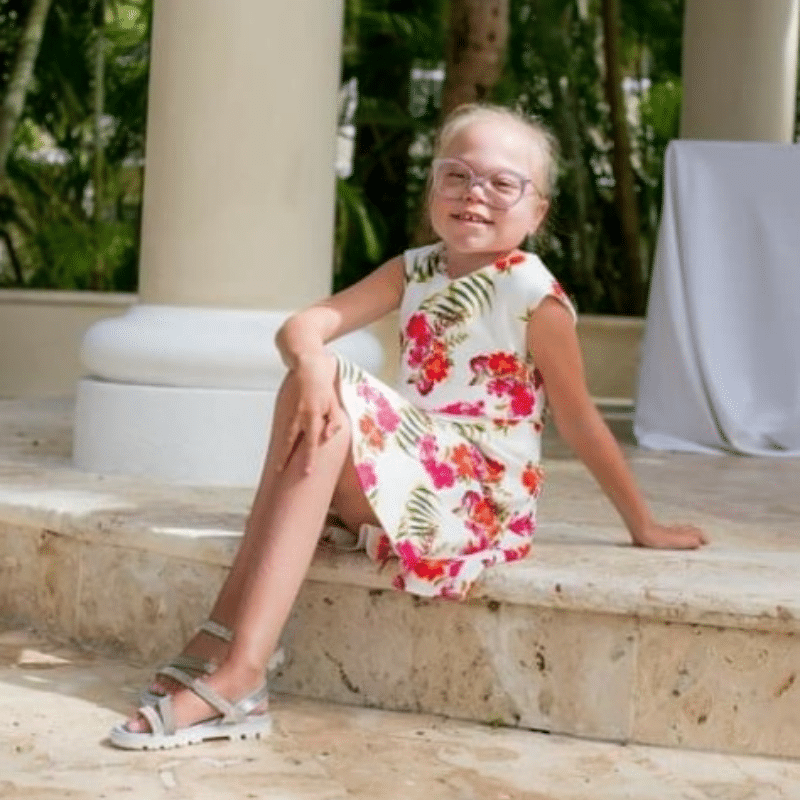
My name is Alessia and I am 11 years old. I live in the UK London; I like dance and art.
At the age of 3 I was diagnosed with Sialidosis after many tests and analyzes. At the age of 8, I was also diagnosed with epilepsy, starting treatment with Kepra.
My health started to deteriorate a year ago after I had Covid-19. I start using a wheelchair for a year and I am totally dependent on my mother or another adult.
Every day I try to enjoy as much as I can every day of my life and I fight with my whole life. I pray every day that God will heal me and give me strength. I hope that soon, we Sialidosis patients will be able to have a treatment that will make our lives easier.

Alexander
A Family’s fight against Sialidosis
Sialidosis is one of the Oligosaccharide family of Lysosomal Storage Diseases. The International Society for Mannosidosis & Related Diseases is proud to present the story of Alexander Skojec and his family’s fight, through the intervention of his father, to raise awareness for this very rare disorder.

Trajano
Hello, my name is Trajano Cerna. I am from Quito-Ecuador, and I live in the United States. I went to School of Medicine for 2 years in Ecuador. I enjoy walking and doing exercises in the park. And I like drinking natural juices. Read more about my journey here.


